Histiocytomas in Dogs: Symptoms, Treatment, and More
Histiocytomas are common skin tumors found in dogs, often appearing as small, red, button-like lumps. They typically occur in younger dogs and can be alarming at first glance.
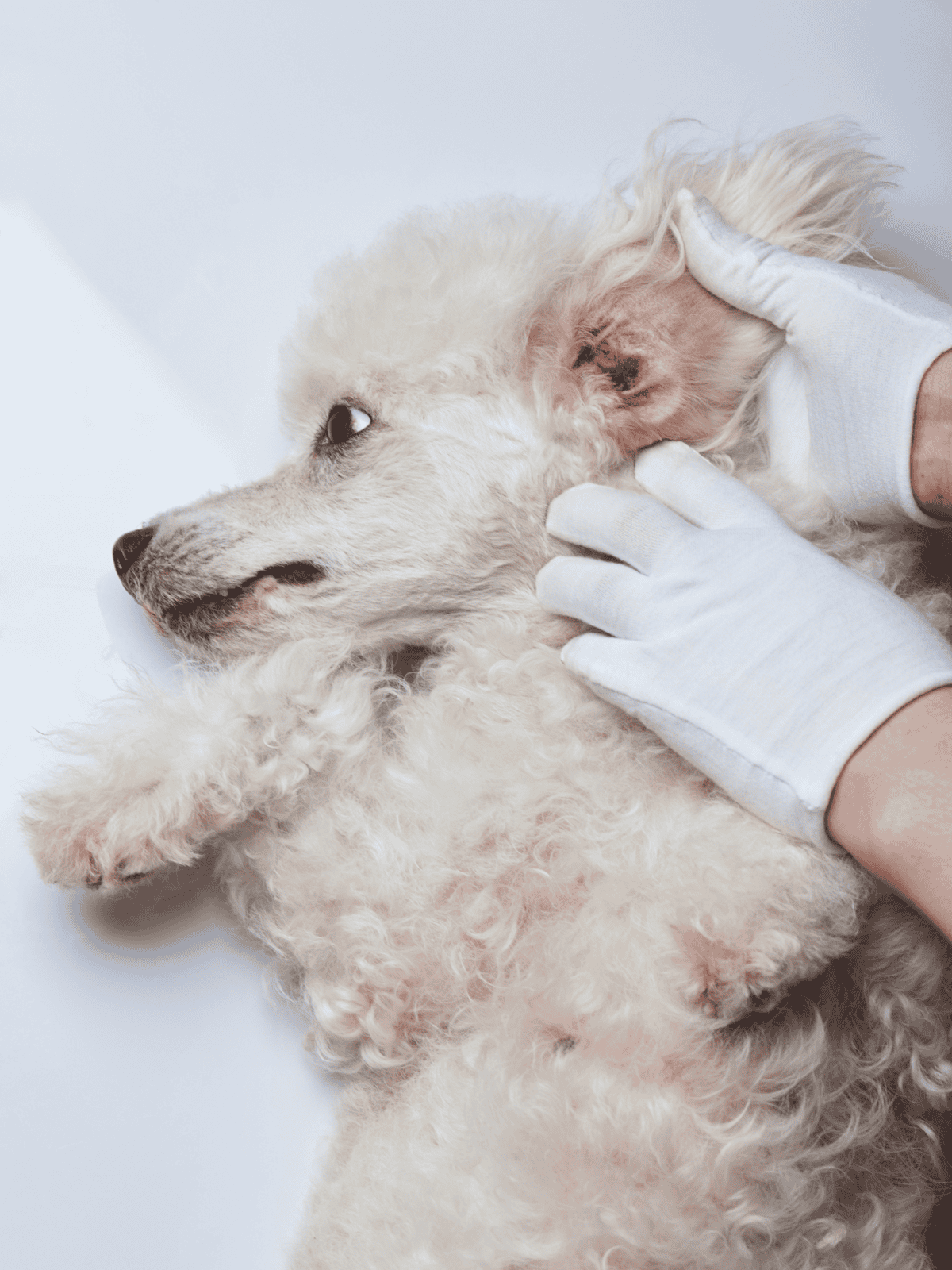
Most histiocytomas are benign and may resolve on their own without treatment. These growths are usually painless and don’t seem to bother the dog. Owners may notice them on the head, neck, or ears, and they rarely spread to other parts of the body.
Understanding when a histiocytoma needs attention is important. While these growths are usually harmless, a vet should check any lump that changes, grows, or causes discomfort. Owners should stay informed about these skin tumors to help keep their pets healthy.
What Are Canine Histiocytomas?
Canine histiocytomas are benign skin growths that are generally not harmful. Often called “button tumors,” they typically appear as single, small red bumps. These lumps are usually less than an inch across and most often grow on a dog’s head, face, ears, or legs.
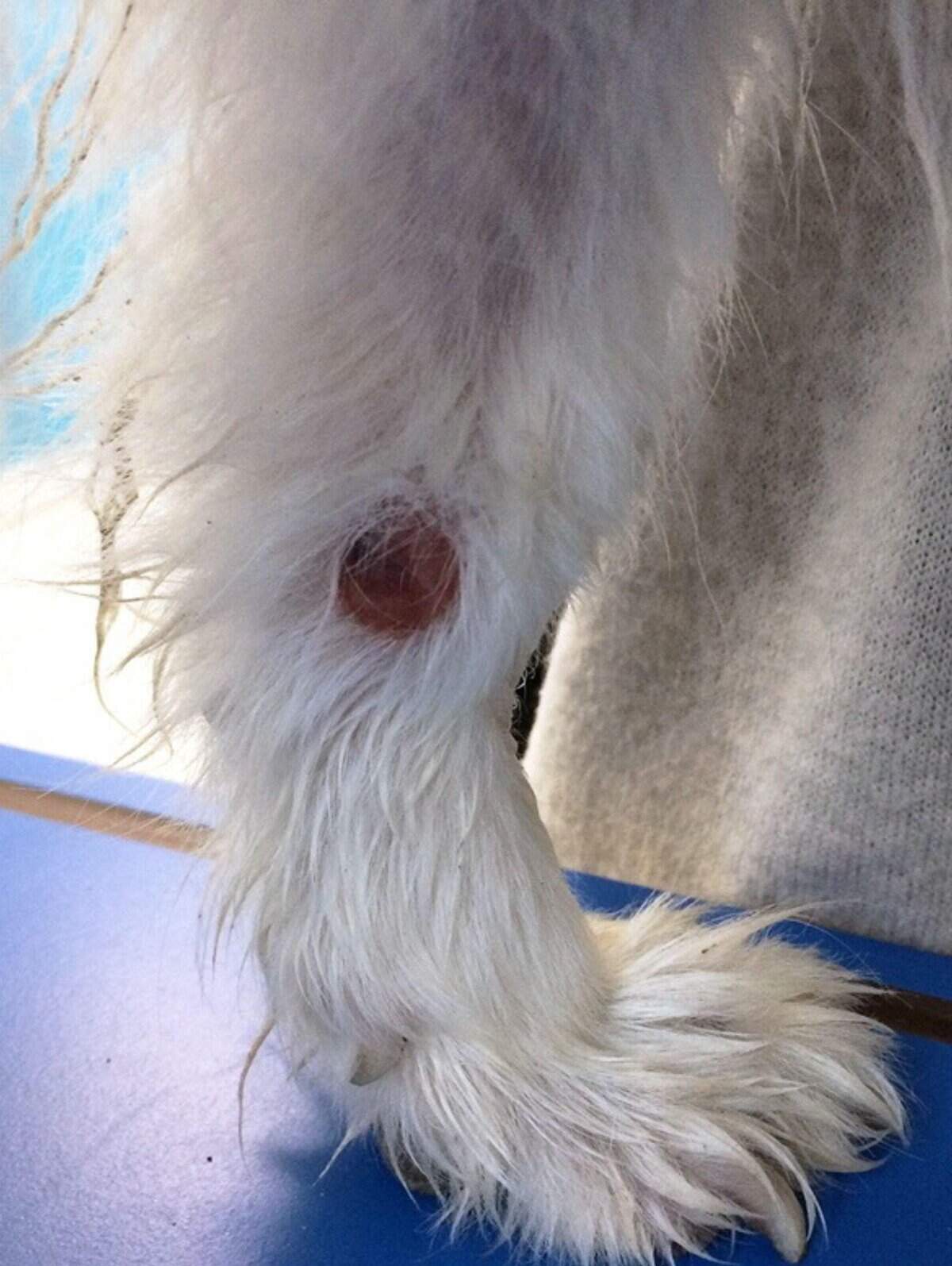
These growths may expand quickly at first but tend to shrink and vanish within a few months. Though they usually have smooth surfaces, they might become sores if the dog licks them too much. It’s important to have a vet examine these lumps to ensure they are not more serious skin issues that look similar.
Signs of Histiocytomas in Dogs
Dogs with histiocytomas might have some noticeable symptoms. These can include a hairless, raised, and red bump on the skin.
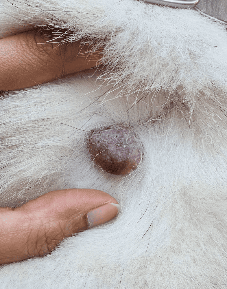
Sometimes, the bump might bleed or cause itching. If there’s an infection, the bump could turn into an open sore with pus or show swelling around it.
Enjoying this read?
We publish this content for free to generate interest in our Premium members' area. By subscribing, you can ask the writer any questions related to pet care and this article, get access to 100+ Premium Pet Care Guides and go Ad-Free with DogFix Premium for $2.99.
Reasons for Histiocytomas in Canines
Histiocytomas in dogs occur due to issues with the immune system. These growths are made of immune cells, so an imbalance in how the immune system works might lead to their formation.
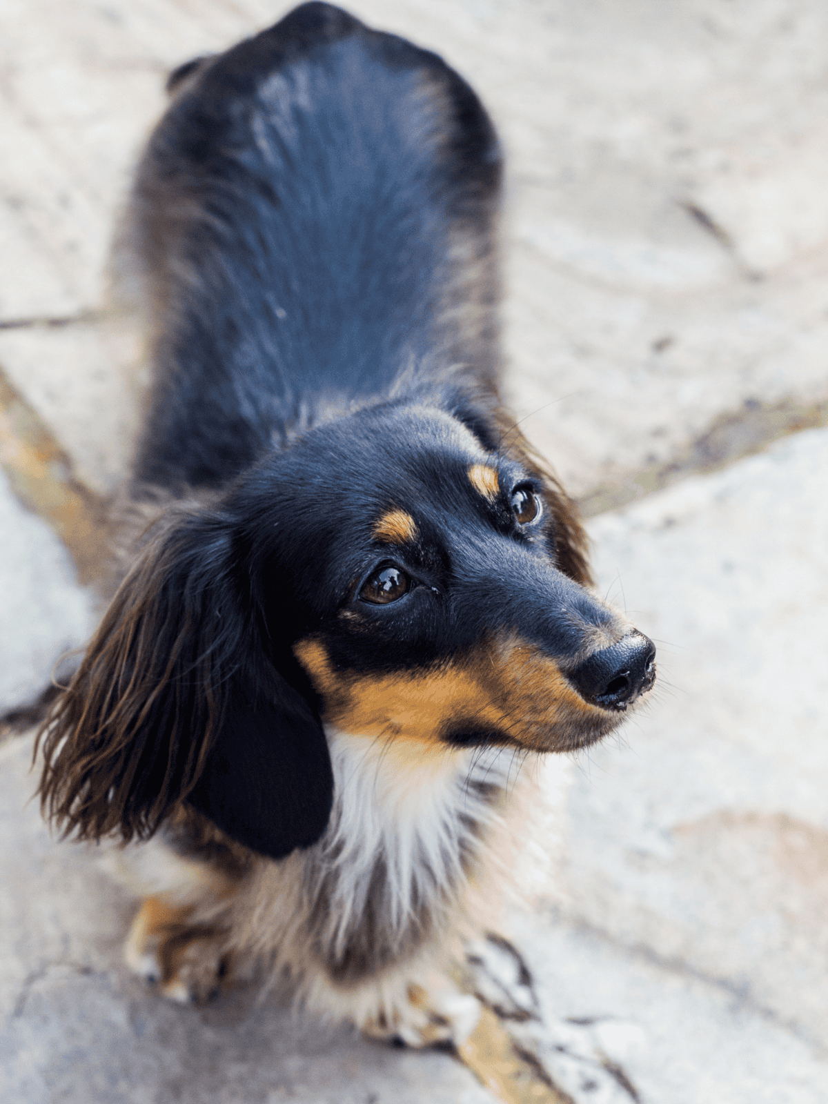
Also, certain breeds seem to get histiocytomas more often, suggesting a genetic link. More research is needed to fully understand these causes. Most histiocytomas occur in younger dogs, typically less than 3 years old. This tumor is more common in certain breeds like Boxers, Bulldogs, and Dachshunds.
How Vets Identify Histiocytomas in Dogs
Veterinarians begin by examining the dog closely, focusing on skin areas. If a suspicious lump is found, further tests are done to determine if it is a histiocytoma.
Physical examination

When a dog shows signs of histiocytoma, a vet will begin with a physical exam. They will check the location and appearance of the lump, which helps them decide on the next steps.
Fine Needle Sample (FNS)
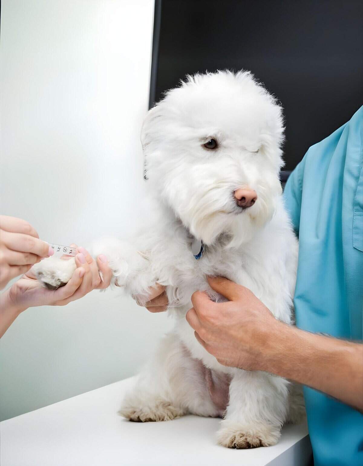
A vet collects a tiny piece of tissue by inserting a thin needle into the lump. This tissue is then stained on a slide and examined under a microscope to check the cell types.
Tissue Sample
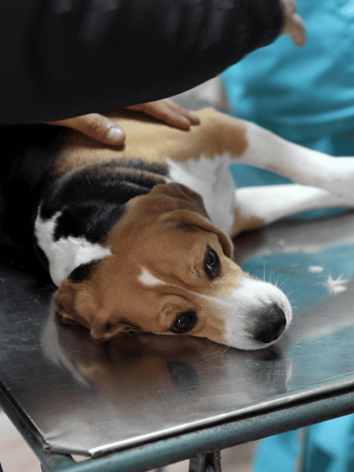
If more detail is needed, a small section or the entire lump might be removed. This sample is sent to a lab for extra tests. This step requires anesthesia or sedation and follows the FNS if the initial results are unclear.
Blood Test
The vet may also suggest blood tests. These tests assess the dog’s overall health and look for any underlying issues.
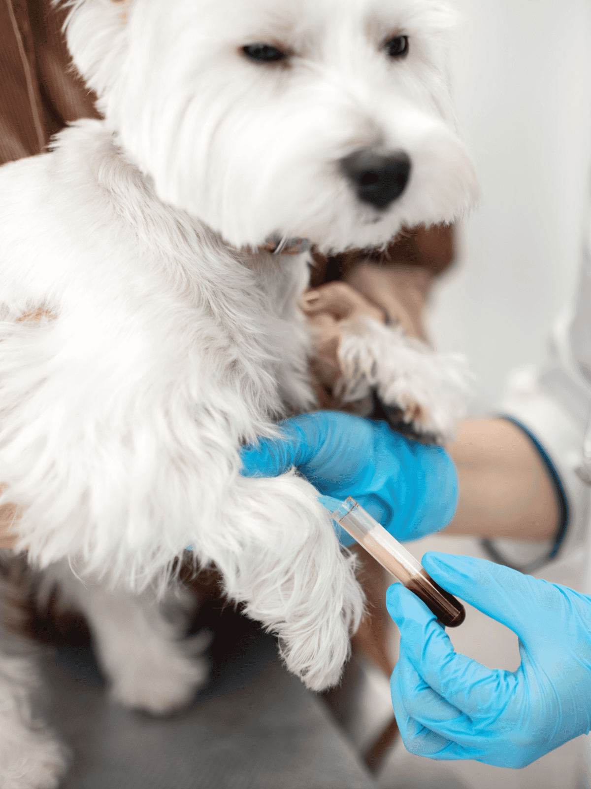
Blood tests can also help determine if the dog is ready for surgery if removal is necessary. Based on these procedures, the vet can create the best plan for managing the histiocytoma.
Treatment Options for Dog Skin Lumps
Most of the time, skin lumps in dogs go away without needing any treatment. The dog’s own immune system often helps shrink these lumps in about three months.
Antibiotic medication
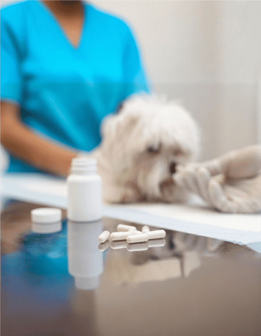
In some cases, especially when the lump is in a spot that gets touched a lot, dogs might lick or scratch it. This can lead to the lump bleeding or getting infected, which is common with lumps on paws. When this happens, using ointments or pills can help treat infections.
Surgery
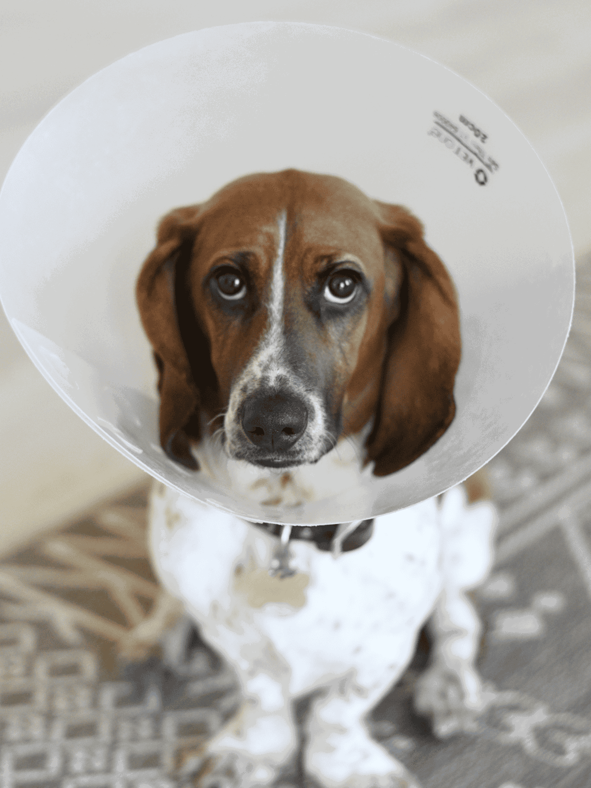
If the lump doesn’t get smaller or bothers the dog, surgery might be the best option. This is usually the last option, as this is an invasive procedure.
Histiocytomas Care and Treatment in Dogs
Histiocytomas in dogs usually don’t bother them, but those on the legs might irritate if they touch the ground. Orthopedic dog beds can help by cushioning these spots.
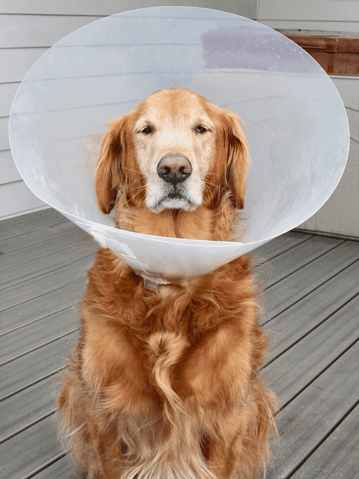
For some dogs, a recovery cone might be needed to stop them from licking or scratching the area. If the histiocytoma starts to bleed or ooze, it’s wise to take the dog to a vet to check for possible infections.
Ways to Prevent Skin Growths in Dogs
Preventing skin growths, such as histiocytomas, can be challenging since their exact cause isn’t fully understood. Annual visits can help detect issues early. Early detection means better treatment outcomes. Check your dog’s skin often. Look for unusual lumps or bumps. Regular grooming sessions make it easier to spot any changes.
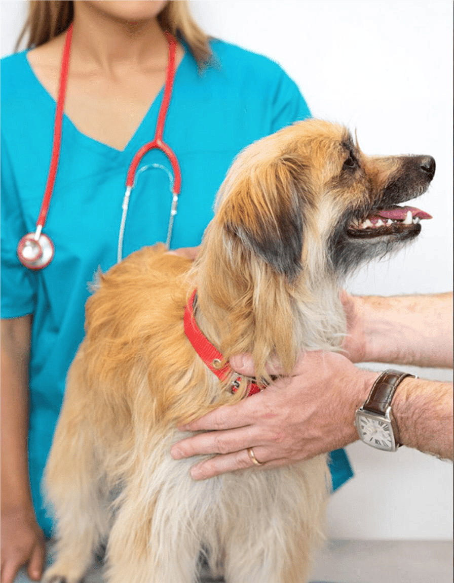
Key steps include keeping an eye on the area to avoid irritation or infection. It’s crucial to take note of any changes and inform the veterinarian right away for advice. By doing this, pet owners can help their dogs remain comfortable and healthy.
When to Consult a Veterinarian
It’s important to know when to seek professional help for your dog. If you notice a new lump or bump, it’s best to get it checked. Histiocytomas often look red, with raised growths. Sometimes, these growths disappear on their own. Monitor the lump for changes in size or colour. If the lump does not improve or seems to grow, consult a veterinarian.
Dogs may show signs of discomfort. If your dog licks, bites, or scratches the growth, it might be painful or itchy. This requires a veterinarian’s attention to prevent infection. Regular check-ups with a veterinarian are important. They ensure your pet remains healthy and issues like histiocytomas are managed promptly.
Common Questions About Histiocytomas in Dogs
Are There Any Home Remedies for Histiocytomas in Dogs?
No home remedies exist for treating histiocytomas in dogs. These growths typically shrink on their own without treatment.
Pet owners should keep a close eye on the dog’s affected skin for any signs of irritation or infection during this period.
How Fast Do Histiocytomas Develop in Dogs?
Histiocytomas grow rapidly within the first month. During this time, they can reach up to an inch in width. After this initial phase of rapid growth, they often begin to regress.
Differentiating Mast Cell Tumors from Histiocytomas in Dogs
Telling mast cell tumors apart from histiocytomas by just looking at them is hard, as they can appear quite similar.
The only way to properly identify these skin masses is by having a veterinarian conduct a fine needle aspiration (FNA) or a biopsy.
